Confocal laser scanning microscopy
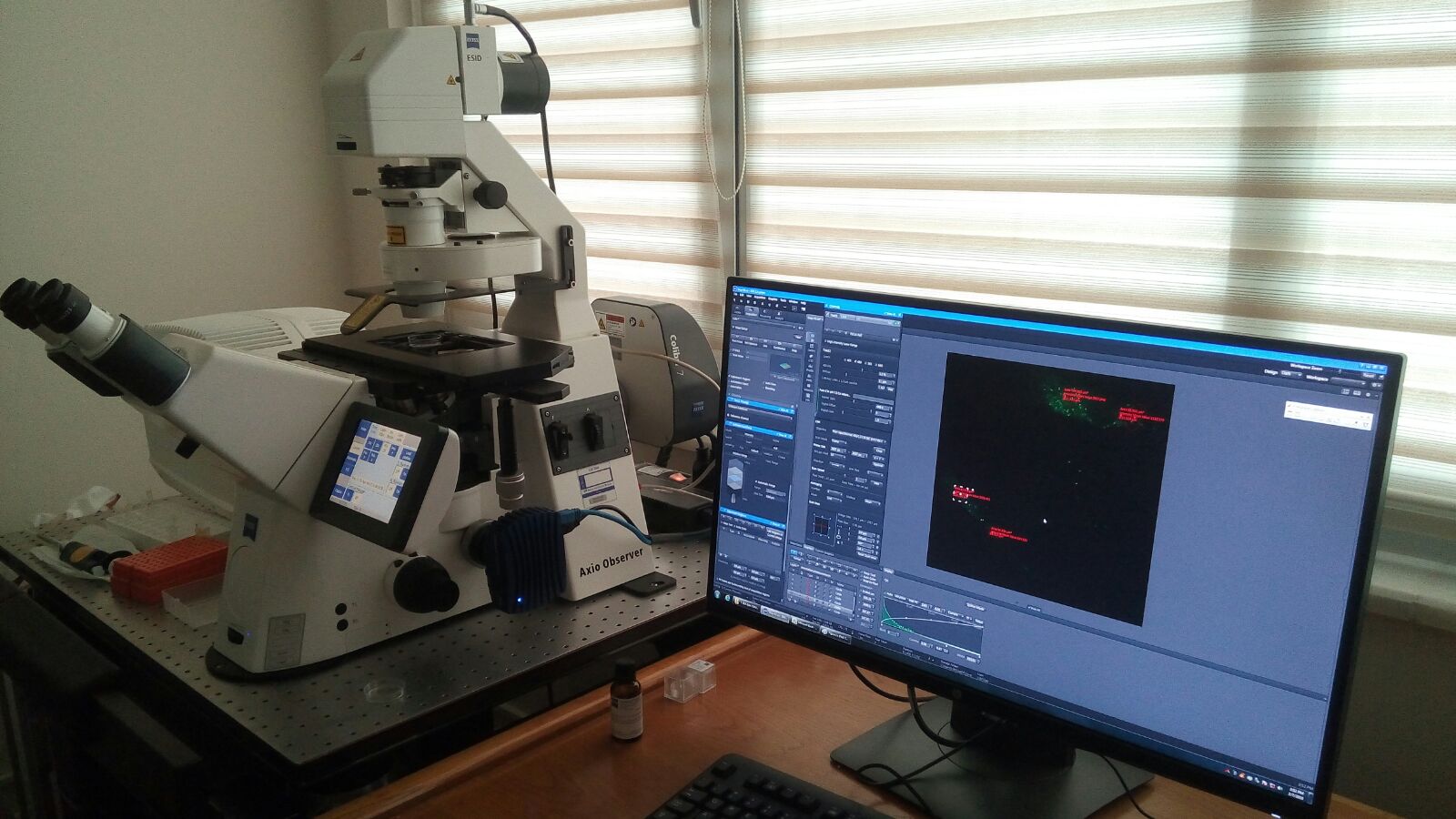
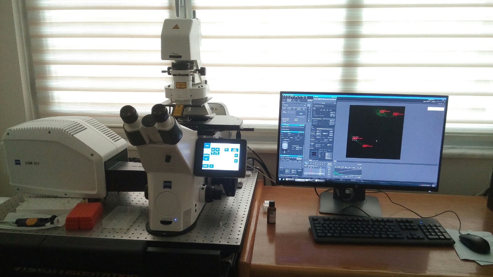
We can better understand structure of cells in fluorescent microscopy by images with high resolution. In confocal laser scanning microscopy we can find out even more. This means that we can view visual sections of tiny structures that would be difficult to physically section and construct 3-D structures from the obtained images. For example, different cells are incubated by different florescent calcium indicator dyes (Fura-2, Flo-3 and Fluo-8) for making visible intracellular free Ca2+. We can also measure the florescent dye concentration as indicator of intracellular free Ca2+ concentration. Basic components of confocal laser scanning microscopy are laser, beam splitter, scanner, objective lens, Z-control, pinhole and photomultiplier tube. In our center, we have modern confocal laser scanning microscopy (Zeiss, LSM800) and we will analyze our neuronal cell samples in the microscope.
Confocal Microskope Course Program
| Lecture 1 | What is the confocal microscope? | Teorik |
| Lecture 2 | Preparation of cells and dyes | Pratik |
| Lecture 3 | Imaging and calculation cytosolic Ca2+ (Fura-2, flo-3 and fluo-8) | Pratik |
| Lecture 4 | Apoptosis analyses (Annexin V and PI) | Pratik |
| Lecture 5 | Mitochondrial membrane depolarization imaging (JC-1) | Pratik |
| Lecture 6 | Cytosolic ROS (Rhodamine 123) imaging and calculation | Pratik |
| Lecture 7 | Cytosolic GSH imaging and calculation | Pratik |
| Lecture 8 | Evaluation of confocal results | Pratik |
| Lecture 9 | How can we prepare the confocal results for a manuscript? | Pratik |
| Lecture 10 | Discussion | Teorik |